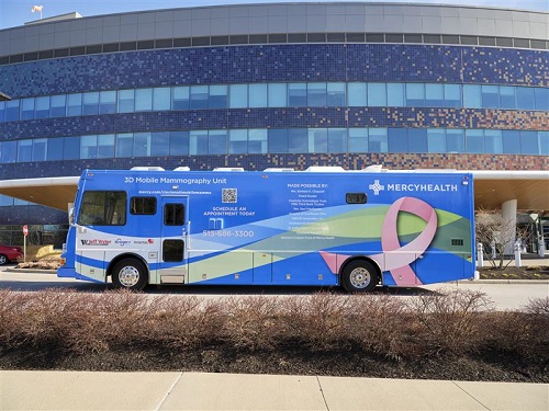
3D Digital Tomosynthesis Mammography
This revolutionary mammography screening generates a series of thin, high-resolution images that allow doctors to view your breast one layer at a time. Using a digital mammography machine with an additional arm that sweeps in an arc over the breast, tomosynthesis creates images that are essentially one-millimeter “slices” of the breast, revealing information that can be hidden in standard digital mammograms. There are many benefits to this state-of-the-art technology, including:
- Improved visualization of breast tissue
- Better ability to detect more and smaller cancers
- Decreased recalls for additional tests
- Less worry for you
Your tomosynthesis exam will include both a 2D and 3D mammogram. For the standard 2D exam, your technologist will take two views of each of your breasts. During the 3D tomosynthesis portion of your exam, the technologist will once again position each of your breasts for two views, but the x-ray arm of the mammography machine will make a quick arc over your breast while taking a series of images at various angles.
Once the exam is complete, the technologist will send your images electronically to the radiologist, who will study them, compare them to previous exams, and then prepare a report of the findings for your physician.
Breast Tomosynthesis in Combination with New C-View Imaging Software
In May 2013 the U.S. Food and Drug Administration (FDA) approved the use of Hologic C-View imaging software. Prior to this decision Hologic’s FDA approved 3D mammography screening exam required an additional conventional 2D mammography image. Consequently, breast tomosynthesis patients were exposed to an increased radiation dose, and slightly increased compression time, compared to a conventional 2D mammography study. C-View imaging software is not a standard feature with a Hologic 3D mammography machine.
Women will benefit from the combination of 3D mammography and C-View software as it eliminates the need for additional radiation exposure required in 3D mammography exams without C-View technology. 3D mammography strengthens the ability of radiologists to find breast cancer and reduces recall rates. This will be of special benefit to women with dense breast tissue.
Screening Mammography
An effective use of mammography as a screening tool is recommended annually for women 40 years of age and older. This exam is used to detect early breast cancer in women who are not experiencing any breast symptoms (pain, lump, nipple discharge, or skin changes).
Diagnostic Mammography
This is a problem-solving mammogram that may be recommended by the radiologist or referring doctor in patients with an abnormal mammogram, breast lump, nipple discharge, or skin changes. This exam is used to help determine the cause of concern in these patients.
 |
Mobile Mammography
Mercy Health also has a Mobile Mammography program. Mercy Health’s three mobile mammography units stop at convenient locations throughout Greater Cincinnati several times each week and offer the latest imaging technology, including tomosynthesis or 3D imaging. Call 513-686-3300 or 1-855-PINK123 (1-855-746-5123) to schedule a mammogram when the mobile mammography unit is at a location convenient to you.
See this heart-warming story about Joretta Kidd, an employee from the Cincinnati Hamilton County Community Action Agency, and Mercy Health Mobile Mammography, which detected her breast cancer early and possibly saved her life.
Find the Mobile Mammography Coach
Quality, Convenient Services
Our specialists strive to deliver the quality you deserve through providing integrated, patient-centered care, coupled with advanced digital mammography technology. We stand out because:
- Vans feature digital mammography, a cutting-edge breast imaging procedure that offers a clearer image, resulting in the most accurate detection of early breast cancer.
- Digital images are reviewed by a physician on the Jewish Hospital Radiology team, giving you the advantage of high-quality, seamless care you can trust.
- Mammograms are reviewed and report made available to the primary care physician within 24 hours.
- Images are conveniently stored electronically and sent by letter or fax through Mercy Health’s electronic medical records system, allowing for immediate consult, which means faster results and greater accessibility.
- We also track digital mammography screenings through a Mammography Reporting System, used to encourage regular screenings.
- The Women’s Services Van uses the R2 ImageChecker, a computer-aided detection system that detects 23.4 percent more breast cancers than mammography alone.
- All three vans meet the standards of the American College of Radiology and the Mammography Quality Standards Act.
Preparation for mammography
- Do not wear deodorant or powder under the arms the day of the procedure.
- When possible, obtain old exams and make them available to the facility where your current exam is being performed.
- Describe any current symptoms to the technologist performing the exam: describe any pain, lumps, discharge, or skin changes.
- Ask when your results will be available and how they will be communicated to you.
